Posted by slomotionshoes | 10 November, 2014
Metatarsal Stress Fracture: Diagnosis

Meeting with a physician is key for early diagnosis and treatment of a metatarsal stress fracture. In many cases patients meet with physicians and report not having a traumatic injury or even increase in activity over the period of time pain becomes apparent. Injury to the metatarsals can be very subtle as day’s walking in poorly supportive shoes like loafers or the minimalist shoes previously discussed. One early sign of a metatarsal stress fracture is swelling to the top of the forefoot with pain that worsens through the day’s activities. Palpable pain to the metatarsal shaft is always noted during medical examination. Manually localizing the pain to the metatarsal associated with the forefoot swelling is key for diagnosis of this injury and can be more effective than x-rays.
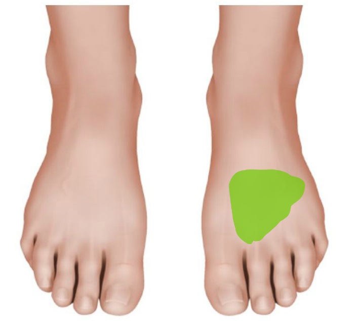
Area of forefoot swelling to the injured foot
X-ray examination will only begin to locate the stress fracture after at least two weeks of injury with first notable periosteal reaction (formation of new bone) and cortical irregularities (hairline cracks to “hard” layer of bone) matching the point of paint and tenderness.
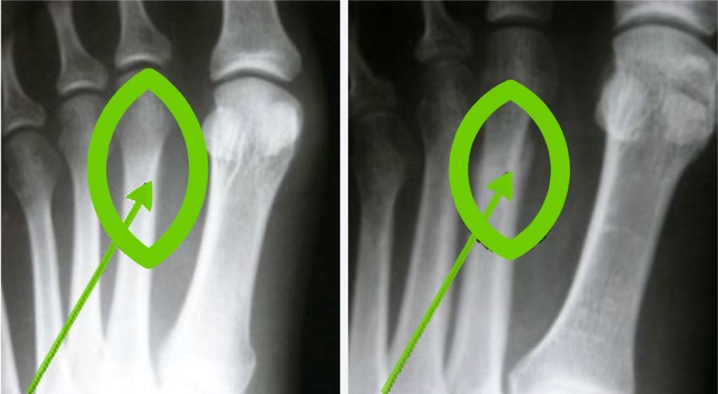
Negative x-ray 2 weeks post injury despite clinical symptoms (left) compared to same patient’s x-ray 3 weeks post injury finally showing signs of stress fracture in the form of new bone growth (right).
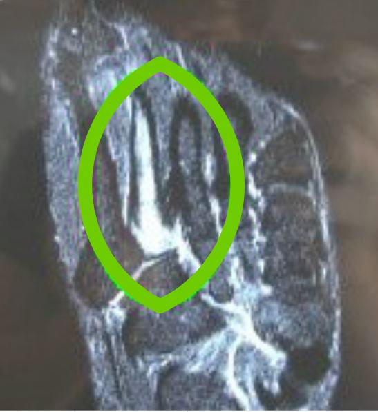
The most effective medical imaging for metatarsal stress fractures diagnosis are bone scans and MRIs as they have high sensitivity to irregularities but are used selectively based on the situation. Bone scans and MRIs are often reserved for high level athletes if there are any question of initial diagnosis. Below is an MRI of a second metatarsal stress fracture showing fluid intensity helping support the diagnosis.